Anterior vs Lateral Corticospinal Tract: Key Differences Explained
The anterior and lateral corticospinal tract represent crucial pathways in our nervous system that control voluntary movement. Have you ever wondered how your brain communicates with your muscles to create precise, intentional movements? The answer lies in these fascinating neural highways. In this comprehensive guide, we'll explore the significant differences between these two tracts and understand why they're so vital for everyday function.
When I first began studying neuroscience, I found the various neural pathways confusing. However, understanding the distinct roles of these corticospinal tracts helped clarify how our brain achieves such remarkable control over our body. The corticospinal tracts are collections of axons carrying signals from the cerebral cortex to the spinal cord, enabling voluntary muscle movements that we often take for granted.
What is the Corticospinal Tract?
Before diving into the differences, let's establish what these tracts actually are. The corticospinal tract (sometimes called the pyramidal tract) is the largest descending pathway found in humans. It's essentially a bundle of nerve fibers that carries movement-related information from the motor cortex in the brain down to the spinal cord.
Think of the corticospinal tract as an information superhighway, transmitting crucial signals that allow you to perform everything from typing on a keyboard to maintaining your posture while sitting. These signals originate in the motor regions of the cerebral cortex and travel through specific pathways to reach their target muscles. The tract gets its name from its origin (cortex) and destination (spinal cord).
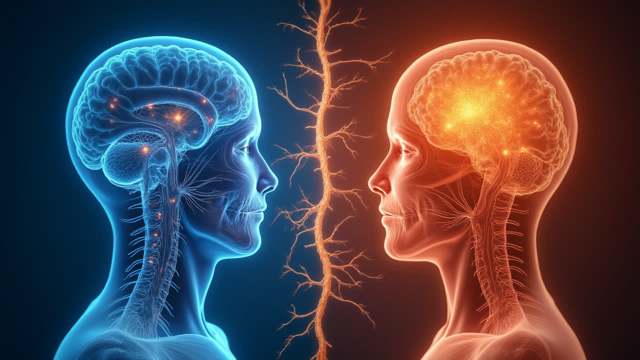
Interestingly, the corticospinal tract isn't a single uniform structure but divides into two main components: the anterior (or ventral) corticospinal tract and the lateral corticospinal tract. Each has distinct characteristics and functions that we'll explore in detail. I've always found it fascinating how these parallel pathways evolved to control different aspects of our movement.
Anterior Corticospinal Tract: Structure and Function
The anterior corticospinal tract, also known as the ventral corticospinal tract or direct pyramidal tract, is a smaller component of the overall system. This tract contains approximately 10% of the nerve fibers within the corticospinal system. One of its most distinguishing features is that it's located near the anterior median fissure of the spinal cord.
Structurally, the anterior tract has some unique characteristics. It primarily exists in the upper regions of the spinal cord and gradually diminishes in size as it descends. In fact, it typically ends around the middle of the thoracic region, meaning it doesn't extend throughout the entire spinal cord. The fibers of this tract notably don't cross at the level of the medulla oblongata in the brain (where the lateral tract fibers cross). Instead, they cross later at the level where they synapse with lower motor neurons.
Functionally, the anterior corticospinal tract specializes in controlling the voluntary movements of axial muscles - those muscles that stabilize and move your trunk. This includes muscles that help maintain posture and perform certain neck and back movements. I've noticed that when explaining this to students, it helps to think of the anterior tract as primarily responsible for keeping you upright and stable.
The anterior tract plays a critical role in posture adjustment and the control of proximal muscles. These are the muscles closer to the midline of your body that help with stability and larger movement patterns rather than fine motor skills. Without this tract functioning properly, maintaining an upright posture would be significantly more difficult.
Lateral Corticospinal Tract: Structure and Function
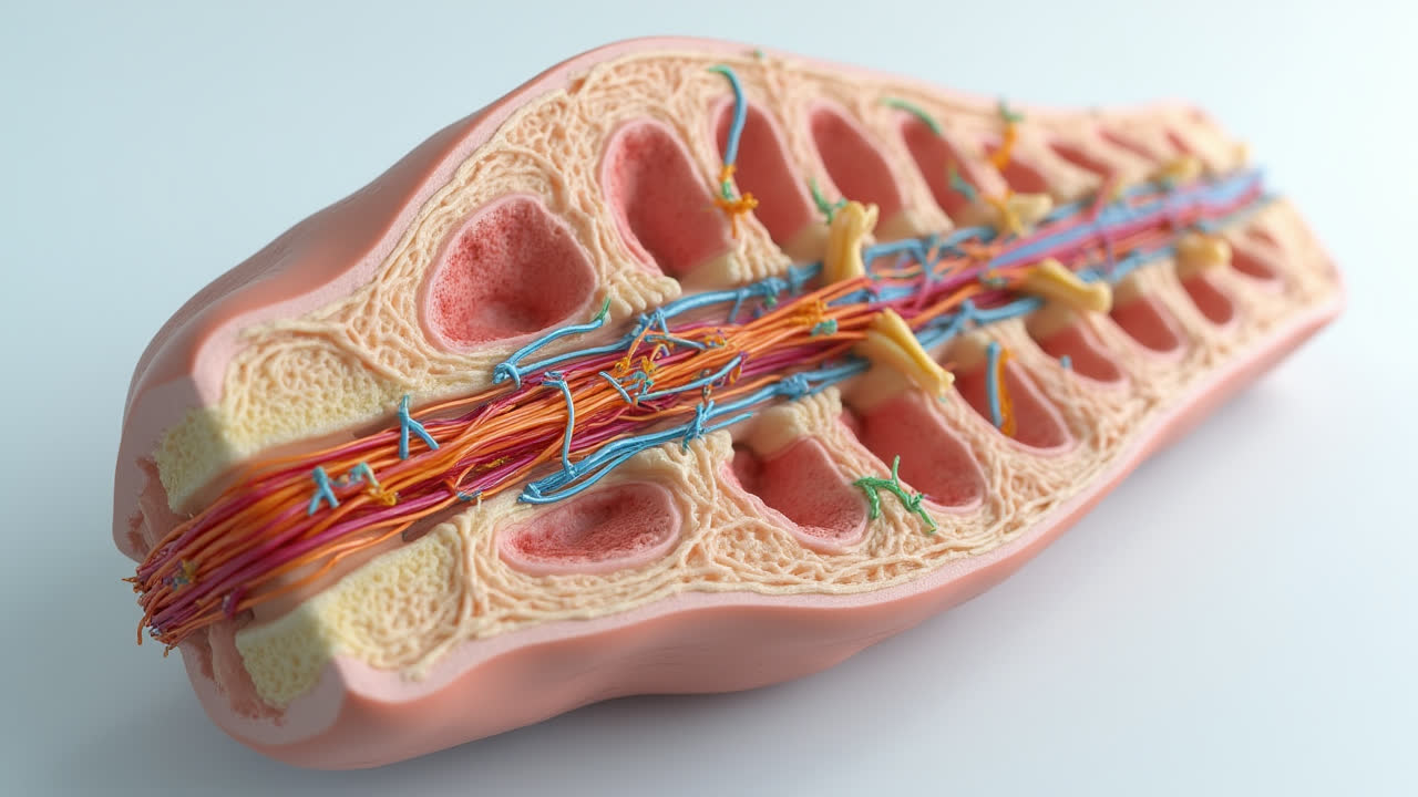
The lateral corticospinal tract is by far the more substantial component, containing approximately 90% of the nerve fibers in the corticospinal system. It's also known as the crossed pyramidal tract or lateral cerebrospinal fasciculus. Unlike its anterior counterpart, the lateral tract extends through the entire length of the spinal cord.
When viewed in cross-section, the lateral corticospinal tract appears as an oval area in the front of the posterior column of the spinal cord, medial to the posterior spinocerebellar tract. Its organization is particularly interesting - in the cervical and lumbar enlargements of the spinal cord (areas serving the arms and legs), this tract is notably larger, reflecting the greater motor control needed for our limbs.
A defining characteristic of the lateral corticospinal tract is that its fibers cross (or decussate) at the level of the medulla oblongata. This crossing explains why the left side of your brain controls the right side of your body, and vice versa - something I've always found to be one of the most fascinating aspects of neuroscience! After crossing, these fibers continue down the opposite side of the spinal cord from their origin in the brain.
The primary function of the lateral corticospinal tract is controlling voluntary movements of the extremity muscles - your arms, hands, legs, and feet. This tract is especially crucial for fine, precise movements like writing, playing instruments, or delicate manipulation tasks. The high proportion of fibers dedicated to this tract underscores the importance of fine motor control in human function and evolution.
I've found that patients with damage to the lateral corticospinal tract often struggle with precision movements and may experience weakness in their limbs. This clinical observation highlights the tract's importance in everyday activities that require dexterity and fine control.
Key Differences Between Anterior and Lateral Corticospinal Tracts
| Feature | Anterior Corticospinal Tract | Lateral Corticospinal Tract |
|---|---|---|
| Proportion of Fibers | Contains 10% of corticospinal fibers | Contains 90% of corticospinal fibers |
| Alternative Names | Ventral corticospinal tract, Direct pyramidal tract | Crossed pyramidal tract, Lateral cerebrospinal fasciculus |
| Extent in Spinal Cord | Ends at mid-thoracic region | Extends through entire spinal cord |
| Decussation Point | At segmental level (later) | At medulla oblongata (earlier) |
| Position in Spinal Cord | Near anterior median fissure | In lateral funiculus, anterior to posterior column |
| Primary Function | Controls axial/trunk muscles | Controls limb/extremity muscles |
| Type of Movement | Posture, proximal muscle control | Fine motor control, precise movements |
| Clinical Significance | Damage affects posture and trunk stability | Damage affects limb coordination and dexterity |
Similarities Between the Tracts
Despite their differences, both the anterior and lateral corticospinal tracts share important common features. They both originate from neurons in the cerebral cortex, specifically from the precentral gyrus (primary motor cortex) and surrounding areas. Both tracts consist of upper motor neurons whose axons form these pathways.
Both tracts are part of the pyramidal system, named for the pyramid-shaped structures they form in the medulla oblongata. They both carry signals related to voluntary movement, distinguishing them from other neural pathways that may control involuntary or reflexive movements. Additionally, both tracts ultimately synapse with lower motor neurons in the spinal cord, which then directly connect to muscles.
Another similarity is that both tracts can be affected by similar conditions. Damage to either tract through stroke, trauma, or degenerative diseases can result in motor deficits. However, the specific symptoms will vary depending on which tract is primarily affected. Understanding these similarities helps provide context for the important functional differences between these parallel motor pathways.
Clinical Significance
The distinct functions of these tracts have important clinical implications. Damage to the corticospinal tracts can occur due to various conditions including stroke, trauma, multiple sclerosis, spinal cord injury, or tumors. When such damage occurs, the resulting symptoms often reflect which tract has been affected.
Lesions affecting the lateral corticospinal tract typically result in contralateral weakness or paralysis of the limbs, particularly affecting fine motor movements. This condition is often accompanied by increased muscle tone (spasticity) and hyperactive reflexes below the level of the lesion. You might see patients struggling with tasks requiring dexterity, such as buttoning a shirt or writing.
In contrast, damage to the anterior corticospinal tract primarily affects trunk stability and postural control. Patients might have difficulty maintaining an upright posture or coordinating movements that require trunk stability. This type of damage is less commonly seen in isolation, as injuries often affect multiple tracts simultaneously.
Interestingly, the body has some capacity for neuroplasticity following damage to these tracts. In some cases, particularly with younger patients, other neural pathways may partially compensate for damaged corticospinal tracts. This offers hope for recovery through intensive rehabilitation and therapeutic interventions.
Evolutionary Perspective
From an evolutionary standpoint, the development of the corticospinal tracts represents a significant advancement in motor control. The lateral corticospinal tract, with its emphasis on fine motor control, is particularly well-developed in humans compared to other mammals. This reflects the evolutionary importance of precise hand movements for tool use and manipulation in human evolution.
The anterior corticospinal tract's focus on axial muscle control aligns with the evolutionary challenges of bipedalism. As humans evolved to walk upright, sophisticated control of trunk muscles became essential for maintaining balance and posture. The differentiation between these two tracts likely represents an evolutionary adaptation that allowed for both stable posture and precise manipulation - two defining features of human movement.
I've often pondered how these specialized neural pathways contributed to human technological and cultural development. Without the fine motor control afforded by the lateral corticospinal tract, activities like writing, crafting tools, and creating art would be impossible. Similarly, without the postural control provided by the anterior tract, the physical stability needed for these activities would be compromised.
FAQs About Corticospinal Tracts
How does damage to the lateral corticospinal tract affect movement?
Damage to the lateral corticospinal tract typically results in contralateral weakness or paralysis, primarily affecting the limbs and fine motor movements. Patients often experience difficulty with precise movements like writing or buttoning clothes. This damage frequently leads to increased muscle tone (spasticity), hyperactive reflexes, and may cause an upgoing Babinski sign - a classic indication of upper motor neuron damage. Recovery depends on the extent of damage and may involve rehabilitation to activate neuroplasticity mechanisms.
Why do the fibers in the lateral corticospinal tract cross at the medulla oblongata?
The crossing (decussation) of fibers at the medulla oblongata explains why the left hemisphere of the brain controls the right side of the body and vice versa. This crossing likely evolved as a solution to the bilateral symmetry of vertebrate bodies. While the evolutionary advantage isn't entirely clear, this arrangement allows for efficient neural processing and integration of sensory and motor information. The decussation occurs during embryonic development through complex molecular signaling processes that guide the growing axons to cross the midline.
Can the brain compensate for damage to one of the corticospinal tracts?
Yes, the brain can partially compensate for damage to corticospinal tracts through neuroplasticity - the brain's ability to reorganize itself by forming new neural connections. This compensation is generally more effective in younger patients and with smaller, partial lesions. The brain may recruit alternative motor pathways, such as the rubrospinal or reticulospinal tracts, to assume some functions of the damaged corticospinal fibers. Intensive rehabilitation therapy can enhance this natural compensatory process by encouraging the formation of new neural circuits, potentially leading to significant functional recovery even after substantial damage.
Conclusion
The anterior and lateral corticospinal tracts represent a fascinating example of functional specialization within the nervous system. While they share a common origin and overall purpose in controlling voluntary movement, their distinct characteristics enable them to serve complementary functions.
The anterior corticospinal tract, with its focus on axial muscle control, ensures we maintain proper posture and trunk stability. Meanwhile, the lateral corticospinal tract, with its extensive fiber network, provides the fine motor control that makes human dexterity possible. Together, these pathways form an integrated system that allows for the remarkable range of movements humans can perform.
Understanding these neural pathways has significant implications for clinical practice, rehabilitation science, and our appreciation of human evolution. As research continues to uncover more details about these complex systems, our ability to address movement disorders and recover from neurological damage will continue to improve.
The next time you perform a complex movement - whether it's maintaining your balance while walking or typing on a keyboard - take a moment to appreciate the incredible neural pathways that make such actions possible. Your corticospinal tracts are silent heroes, working continuously to translate your intentions into physical reality.