Cerebrum vs Cerebral Cortex: Understanding Key Differences & Functions
Cerebrum vs Cerebral Cortex: Key Differences & Functions
The human brain is a marvel of biological engineering, containing roughly 86 billion neurons that form trillions of connections. Within this complex organ, the cerebrum and cerebral cortex play vital roles in everything from conscious thought to voluntary movement. But what exactly distinguishes these two crucial brain structures? How do they relate to each other, and what specific functions does each one perform?
Many people confuse the cerebrum and cerebral cortex or use the terms interchangeably, but they're actually distinct parts with unique characteristics. The cerebrum constitutes the largest portion of the brain, while the cerebral cortex specifically refers to the outer layer of the cerebrum. Understanding these differences isn't just academic—it helps us comprehend how our brain processes information, controls our body, and creates our experience of consciousness.
In this comprehensive guide, we'll explore the relationship between the cerebrum and cerebral cortex, examining their structure, composition, and functions. Whether you're a student of neuroscience, a healthcare professional, or simply curious about the inner workings of the brain, this article will provide clear explanations of these fascinating brain components. Let's dive into the intricate world of neuroanatomy!
What is the Cerebrum?
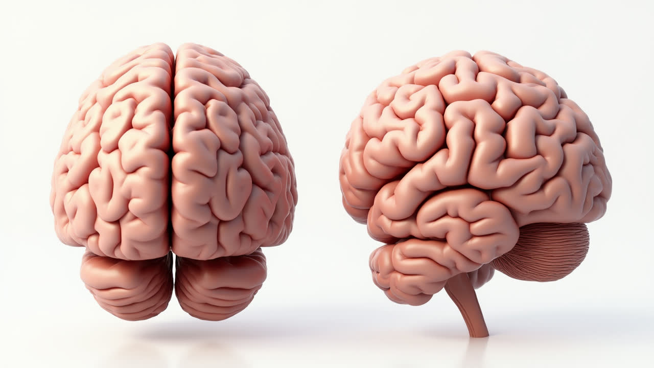
The cerebrum is the largest and most prominent part of the human brain, accounting for approximately 80% of the brain's total weight. Located in the anterior portion of the skull, this massive structure dominates the brain's appearance and is responsible for most higher cognitive functions. When most people picture a brain, they're actually visualizing the cerebrum with its characteristic walnut-like appearance.
Anatomically, the cerebrum consists of two hemispheres—left and right—separated by a deep groove called the longitudinal fissure. These hemispheres are connected by a thick bundle of nerve fibers known as the corpus callosum, which allows the two sides to communicate with each other. Each hemisphere is further divided into four lobes: the frontal, parietal, temporal, and occipital lobes. These divisions aren't merely arbitrary—each lobe specializes in different functions and processes.
The cerebrum contains both gray and white matter. The gray matter consists primarily of neuronal cell bodies and dendrites, while white matter is composed of myelinated axons that connect different parts of the brain. This distinction is crucial for understanding how the cerebrum processes and transmits information throughout the nervous system. The outer layer of the cerebrum is the cerebral cortex (which we'll discuss in more detail below), composed primarily of gray matter, while the interior regions contain the white matter.
Functionally, the cerebrum controls voluntary movements, processes sensory information, and handles complex cognitive tasks including reasoning, learning, memory, language, and emotional processing. The left hemisphere typically specializes in analytical and verbal tasks, while the right hemisphere excels at spatial relationships and pattern recognition. However, this specialization isn't absolute—both hemispheres work together for most complex tasks.
Interestingly, the cerebrum exhibits contralateral control, meaning the right hemisphere controls the left side of the body, and the left hemisphere controls the right side. This crossing of neural pathways occurs in the brainstem and is a fundamental feature of the vertebrate nervous system. Damage to one hemisphere often results in deficits on the opposite side of the body, a fact that has helped neurologists map brain functions through studying patients with localized brain injuries.
What is the Cerebral Cortex?
The cerebral cortex is the outermost layer of the cerebrum, composed of folded gray matter that ranges from 2 to 5 millimeters in thickness. This wrinkled, convoluted surface is perhaps the most distinctive feature of the human brain and is often what gives the brain its characteristic appearance. If you were to look at a cross-section of the brain, the cerebral cortex would appear as the darker outer rim surrounding the lighter white matter below.
The folded structure of the cerebral cortex is one of its most remarkable features. These folds, consisting of ridges (gyri) and grooves (sulci), dramatically increase the surface area of the cortex without requiring a proportionally larger skull. In fact, if you were to flatten out the human cerebral cortex, it would cover an area of about 2.5 square feet—roughly the size of a large dinner napkin! This extensive surface area allows for the tremendous neural processing power that characterizes human cognition.
Microscopically, the cerebral cortex contains an estimated 16 billion neurons, organized into six distinct layers with different cell types and connections. These neurons form incredibly complex networks that process and integrate information. Unlike the white matter of the cerebrum's interior, the gray matter of the cerebral cortex consists primarily of neuronal cell bodies, dendrites, and unmyelinated axons, giving it its characteristic grayish appearance in preserved specimens.
Functionally, the cerebral cortex is central to our most distinctly human attributes. It's responsible for conscious perception, voluntary movement, language, logical reasoning, planning, and personality. Different regions of the cortex specialize in particular functions—the motor cortex controls voluntary movements, the sensory cortex processes incoming sensory information, the visual cortex interprets visual input, and association areas integrate information from multiple sources to form complex thoughts and memories.
Perhaps most significantly, the cerebral cortex is considered the seat of human consciousness. The intricate processing that occurs within its neural networks gives rise to our subjective experiences, self-awareness, and the ability to reflect on our own thoughts. This makes the cerebral cortex not just a biological structure but a philosophical wonder—the physical substrate of what we experience as mind and consciousness.
Cerebrum vs Cerebral Cortex: Detailed Comparison
| Feature | Cerebrum | Cerebral Cortex |
|---|---|---|
| Definition | The largest part of the brain consisting of two hemispheres | The outer layer of the cerebrum composed of folded gray matter |
| Location | Anterior and superior part of the brain | Outermost layer of the cerebrum |
| Composition | Contains both gray and white matter | Primarily gray matter (neuronal cell bodies and dendrites) |
| Structure | Two hemispheres connected by corpus callosum | Highly folded surface with gyri (ridges) and sulci (grooves) |
| Thickness | Makes up about 80% of total brain volume | Approximately 2-5 millimeters thick |
| Primary Function | Controls voluntary movements and higher cognitive functions | Processes conscious thought, perception, and complex behaviors |
| Regions | Divided into four lobes: frontal, parietal, temporal, and occipital | Contains specialized areas: motor, sensory, and association areas |
| Cellular Arrangement | Varied organization throughout different regions | Organized in six distinct layers with different cell types |
The Key Relationship Between Cerebrum and Cerebral Cortex
Understanding the relationship between the cerebrum and cerebral cortex is crucial for grasping how the brain functions as an integrated whole. The cerebral cortex is not a separate entity from the cerebrum but rather constitutes its outer layer. Think of the cerebrum as an entire apple, and the cerebral cortex as the apple's skin—the cortex forms the outer boundary of the cerebrum, covering its surface and harboring most of its neuronal cell bodies.
This relationship explains why damage to the cerebral cortex often affects functions associated with the cerebrum. When a stroke or injury damages part of the cortex, the corresponding cerebral functions may be impaired because the cortex contains the cell bodies of neurons whose axons extend into deeper regions of the cerebrum. For instance, damage to the motor cortex can result in paralysis or weakness in parts of the body controlled by that region, demonstrating how cortical damage cascades throughout the cerebrum's functionality.
The interaction between the cerebral cortex and the underlying white matter of the cerebrum represents a beautiful example of structural and functional integration in the brain. The cortex processes information and makes decisions, while the white matter fiber tracts of the cerebrum transmit this information to other brain regions and the rest of the body. This partnership allows for both localized processing in specialized cortical areas and distributed processing across broader cerebral networks.
Evolutionarily, the relationship between the cerebrum and cerebral cortex highlights an interesting trend. As mammals evolved increasingly complex behaviors, their cerebral cortices became progressively more folded and intricate, allowing for greater neural processing capability without requiring proportionally larger skulls. Humans, with our remarkably advanced cognitive abilities, have the most highly developed cerebral cortex relative to overall brain size among all animals. This evolutionary emphasis underscores the critical importance of the cortex within the larger cerebral structure.
In developmental terms, the cerebrum and cerebral cortex follow fascinating growth patterns. During embryonic development, the cerebrum begins as a smooth structure, with the characteristic folds of the cerebral cortex developing later in gestation. After birth, the cerebrum continues to grow, with the cerebral cortex undergoing dramatic changes as new neural connections form in response to experience and learning. This developmental plasticity allows the brain to adapt to environmental demands and forms the biological basis for our lifelong capacity to learn and change.
Functions of the Cerebrum and Cerebral Cortex
While we've touched on some functions of both structures, let's delve deeper into the specific roles they play in our daily cognitive and physical experiences. The cerebrum, as the largest part of the brain, handles a wide array of functions. It's the center for voluntary movement, allowing us to walk, write, speak, and perform countless other deliberate actions. It processes and integrates sensory information from our eyes, ears, skin, and other sense organs, giving us our experience of the world around us.
Beyond these basic functions, the cerebrum enables higher cognitive processes like problem-solving, decision-making, planning, and abstract thinking. It's responsible for our personality traits, emotional responses, and social behaviors. Memory formation and retrieval also depend heavily on cerebral structures, particularly the hippocampus and surrounding regions. Language processing—including understanding, producing, and manipulating language—is another sophisticated function of the cerebrum, primarily localized in the left hemisphere for most people.
The cerebral cortex, as the information processing center of the brain, performs even more specialized functions within its different regions. The primary motor cortex, located in the frontal lobe, controls voluntary movements with remarkable precision. The primary somatosensory cortex, situated in the parietal lobe, processes touch, temperature, and pain sensations from throughout the body. Visual information is processed in the occipital lobe, while auditory signals are handled by regions in the temporal lobe.
Association areas of the cerebral cortex perform some of the most complex functions. The prefrontal cortex, for instance, handles executive functions like planning, impulse control, and social judgment. Broca's and Wernicke's areas specialize in language production and comprehension, respectively. The association cortices integrate information from multiple sensory modalities, allowing us to form unified perceptions from diverse inputs—for example, recognizing an apple through its appearance, smell, taste, and texture simultaneously.
Perhaps most fascinatingly, the cerebral cortex is intimately linked with consciousness itself. When you're aware of your surroundings, thinking about your future, or reflecting on who you are as a person, you're engaging your cerebral cortex. It's not just responsible for what we think, but for the fact that we can think about our thinking—a meta-cognitive ability that may be uniquely human. Damage to the cortex can result in alterations to consciousness, from subtle changes in perception to complete loss of awareness in cases of severe injury.
Clinical Significance of Understanding the Difference
The distinction between the cerebrum and cerebral cortex has significant implications in clinical neurology and neurosurgery. When physicians diagnose and treat brain conditions, precisely locating the affected area—whether it's in the cerebral cortex or deeper cerebral structures—can dramatically impact treatment approaches and prognosis. For instance, cortical lesions (damage to the cerebral cortex) often produce different symptoms than damage to subcortical structures within the cerebrum.
Stroke, one of the leading causes of disability worldwide, provides a clear example of this clinical relevance. A cortical stroke affects the outer layer of the cerebrum and typically results in specific deficits related to the functions of the damaged cortical region—perhaps speech difficulties if Broca's area is affected, or visual field defects if the occipital cortex is damaged. In contrast, subcortical strokes affecting white matter tracts deeper in the cerebrum often cause more widespread dysfunction, as these pathways connect multiple brain regions.
Neurosurgeons planning operations must carefully distinguish between cortical and deeper cerebral structures. Some surgeries, like those for certain types of epilepsy, may involve removing small portions of the cerebral cortex while preserving underlying white matter tracts. Other procedures, such as deep brain stimulation for Parkinson's disease, target structures deep within the cerebrum, requiring surgeons to navigate past the cerebral cortex without damaging it. These delicate operations require precise knowledge of cerebral and cortical anatomy.
In neuroimaging, technologies like MRI and CT scans allow physicians to visualize the cerebrum and cerebral cortex non-invasively. Functional MRI (fMRI) can even show which parts of the cerebral cortex are active during specific tasks, revolutionizing our understanding of brain function and providing valuable diagnostic information. Differentiating between cortical atrophy (shrinkage of the cerebral cortex) and more general cerebral atrophy helps in diagnosing conditions like Alzheimer's disease versus other forms of dementia.
Understanding the relationship between the cerebrum and cerebral cortex also informs rehabilitation strategies for brain injuries. Because different regions of the cerebral cortex have some capacity for taking over functions from damaged areas (neuroplasticity), targeted rehabilitation exercises can help patients recover lost abilities by retraining intact cortical regions. The more precisely a clinician can identify which cortical areas remain functional after injury, the more effectively rehabilitation can be tailored to the individual patient.
Conclusion: The Remarkable Interplay of Brain Structures
The relationship between the cerebrum and cerebral cortex exemplifies the beautiful complexity of the human brain. While the cerebrum encompasses the largest portion of our brain with its dual hemispheres and internal white matter, the cerebral cortex forms its critical outer layer where much of our conscious experience is processed. Together, these structures enable everything from basic sensory processing to our most sophisticated cognitive abilities.
Understanding the distinction between these structures isn't merely an academic exercise—it provides essential insights into how our brain functions, how it can malfunction in disease, and how we might better treat neurological conditions. As neuroscience continues to advance, our knowledge of these remarkable brain structures will undoubtedly deepen, potentially leading to better treatments for brain injuries, developmental disorders, and neurodegenerative diseases.
The next time you ponder a difficult problem, enjoy a beautiful sunset, or engage in conversation with a friend, take a moment to appreciate the incredible work being done by your cerebrum and cerebral cortex. These remarkable structures not only enable you to experience the world but also to contemplate your own experience—a truly extraordinary evolutionary achievement.
Frequently Asked Questions About the Cerebrum and Cerebral Cortex
What happens if the cerebral cortex is damaged?
Damage to the cerebral cortex can cause a wide range of symptoms depending on which specific area is affected. Motor cortex damage may result in weakness or paralysis on the opposite side of the body. Damage to sensory areas might cause numbness or abnormal sensations. Injury to speech areas could lead to language disorders like aphasia. Visual cortex damage often results in specific visual field defects. The effects of cortical damage are usually highly localized and specific to the functions of the affected region, though severe or widespread damage can affect consciousness itself. Unlike some other brain regions, the cerebral cortex has limited ability to regenerate after injury, though neuroplasticity allows for some functional recovery as other areas adapt to compensate for the damage.
How does the cerebrum differ between humans and other mammals?
The human cerebrum stands out among mammals primarily in its size relative to body weight and its degree of cortical folding. While all mammals have a cerebrum divided into two hemispheres, the human cerebrum is proportionally much larger, particularly the frontal lobes associated with planning and higher cognition. Our cerebral cortex is also significantly more folded (convoluted) than that of most other mammals, providing greater surface area for neural processing. These differences reflect our species' evolutionary emphasis on complex social behaviors, tool use, language, and abstract thought. Interestingly, dolphins and some whale species also have highly convoluted cerebral cortices, though their brain architecture is adapted for different environmental challenges than those faced by humans.
Can you live without parts of your cerebrum or cerebral cortex?
Yes, it is possible to survive without certain parts of the cerebrum or cerebral cortex, though the effects vary dramatically depending on which areas are missing. Hemispherectomy—the surgical removal of an entire cerebral hemisphere—is sometimes performed in children with severe epilepsy, and remarkably, many develop nearly normal cognitive function as the remaining hemisphere compensates. Smaller portions of the cerebral cortex are sometimes removed to treat localized epilepsy or tumors, with specific deficits depending on the area removed. The brain's plasticity is greatest in young children, allowing for substantial functional recovery after such procedures. However, complete loss of the cerebrum or cerebral cortex is incompatible with conscious life, though some basic reflexes and autonomic functions controlled by the brainstem might continue.