Metacarpal vs Metatarsal: Key Differences in Hand and Foot Bones
Have you ever wondered about the bones that give structure to your hands and feet? The human skeletal system is fascinatingly complex, with metacarpal and metatarsal bones playing crucial roles in our everyday movements. Though they sound similar and serve comparable purposes, these bones have distinct characteristics that set them apart. Understanding these differences can give us valuable insights into human anatomy and how our bodies function.
When you make a fist, the knuckles that protrude are actually the heads of your metacarpal bones. Similarly, when you stand on tiptoe, you're putting pressure on your metatarsal bones. These seemingly small skeletal components are fundamental to our ability to grasp objects, maintain balance, and walk properly. Let's dive deeper into what makes these bones unique and how they contribute to our daily activities.
What Are Metacarpal Bones?
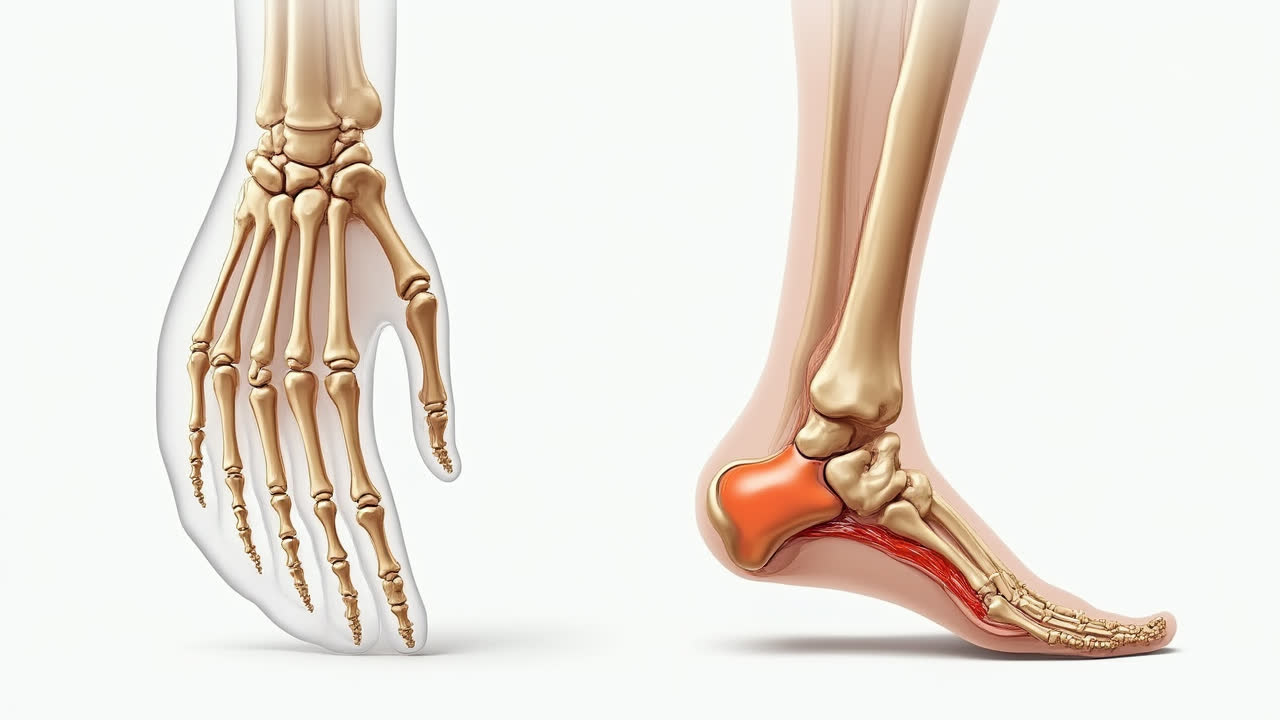
The metacarpal region forms the intermediate part of the hand, connecting the wrist to the fingers. There are five metacarpal bones in each hand, numbered from the thumb side (radial) to the little finger side (ulnar). These bones are classified as long bones despite their relatively small size because they share the same structural characteristics as other long bones in the body.
Each metacarpal consists of three distinct parts: a proximal base that connects to the wrist (carpal) bones, a shaft (or body) that forms the palm, and a distal head that connects to the finger bones (phalanges). The base is typically wider and more irregular in shape, allowing for stable articulation with the carpal bones. The shaft is slightly curved, creating the hollow of the palm, while the head is rounded to facilitate movement at the knuckle joint.
The first metacarpal (thumb) deserves special mention as it's shorter and more mobile than the others. This unique structure enables the thumb's opposability – the ability to touch the tips of other fingers – which is essential for precise grip and manipulation of objects. Without this special adaptation, many of the fine motor skills we take for granted would be impossible. Ever tried picking up a small object without using your thumb? It's remarkably difficult!
Metacarpal bones form two types of important joints. At their proximal ends, they form carpometacarpal joints with the wrist bones, allowing for various movements of the hand. At their distal ends, they create metacarpophalangeal joints (knuckles) with the proximal phalanges of the fingers. These joints are crucial for the flexibility and dexterity that human hands are known for. The metacarpophalangeal joints in particular are condyloid joints, allowing movement in two planes – flexion/extension and abduction/adduction.
What Are Metatarsal Bones?
Moving down to the feet, the metatarsal bones form the middle portion of the foot, connecting the ankle to the toes. Like their hand counterparts, there are five metatarsal bones in each foot, numbered from the big toe (medial) to the little toe (lateral). These bones are essential components of the foot's architectural structure.
Structurally, metatarsals also have three parts: a proximal base that articulates with the tarsal (ankle) bones, a shaft that forms part of the foot's arch, and a distal head that connects to the toe bones. However, there are notable differences in their dimensions and proportions compared to metacarpals. Metatarsals tend to be longer and stronger, as they must support the entire body weight during standing and walking.
The first metatarsal (big toe) is particularly thick and strong compared to the others, reflecting its importance in weight-bearing and propulsion during walking. It's significantly shorter and wider than the second metatarsal, which is typically the longest of the five. This arrangement contributes to the transverse arch of the foot, which distributes weight across the foot and provides stability.
Metatarsal bones form three types of joints: tarsometatarsal joints with the ankle bones, metatarsophalangeal joints with the toes, and intermetatarsal joints with each other. These joints collectively allow for the complex movements needed for walking, running, and maintaining balance. The metatarsophalangeal joint of the big toe is especially important during the push-off phase of gait, where it must withstand forces several times the body weight.
I once had a friend who fractured his fifth metatarsal while dancing at a wedding. He described the pain as surprisingly intense for such a small bone, and his recovery took nearly eight weeks! This personal anecdote highlights just how crucial these bones are to our mobility – when one is injured, it can significantly impact our ability to perform daily activities.
Comparative Analysis: Metacarpal vs Metatarsal
While metacarpals and metatarsals are analogous structures, they have evolved different characteristics to suit their specific functions. Let's break down their key differences and similarities to get a clearer picture of these important skeletal components.
| Feature | Metacarpal Bones | Metatarsal Bones |
|---|---|---|
| Location | Hand, between carpal bones and phalanges | Foot, between tarsal bones and phalanges |
| Number | Five in each hand | Five in each foot |
| Length | Relatively shorter | Longer (especially 2nd metatarsal) |
| Strength | Lighter, more delicate structure | Stronger, built for weight-bearing |
| Joint Types | Carpometacarpal and metacarpophalangeal | Tarsometatarsal, metatarsophalangeal, and intermetatarsal |
| Primary Function | Support for hand dexterity and grip | Weight-bearing and propulsion during walking |
| Most Mobile | First (thumb) metacarpal | First (big toe) metatarsal |
| Common Injuries | Boxer's fracture (5th metacarpal) | Jones fracture (5th metatarsal), stress fractures |
One of the most striking differences between these bone groups relates to their primary functions. Metacarpals are primarily designed for versatility and dexterity, allowing for the precise manipulations that human hands are capable of. In contrast, metatarsals evolved for stability, weight-bearing, and efficient locomotion. These functional differences are reflected in their structural adaptations.
Another interesting difference lies in the mobility patterns. The thumb metacarpal has a unique saddle joint with the trapezium bone, allowing for opposition movement that's crucial for grasping objects. The big toe metatarsal, while important, doesn't have this same degree of mobility – and for good reason! Can you imagine trying to maintain balance if your big toe could move like your thumb? Sometimes, limited mobility is actually advantageous.
Clinical Significance and Common Injuries
Understanding the differences between metacarpals and metatarsals becomes particularly relevant in clinical settings. These bones are susceptible to various injuries and conditions that can significantly impact a person's quality of life.
In the hand, a common injury is the "boxer's fracture," which typically involves the neck of the fifth metacarpal. This injury often results from punching a hard surface with a closed fist. The metacarpal bones can also be affected by arthritis, particularly at the carpometacarpal joint of the thumb, which can cause significant pain and reduced grip strength. Hand surgeons and therapists work extensively with metacarpal issues, developing specialized techniques to restore function.
Metatarsal injuries present different challenges. Stress fractures of the metatarsals are common among athletes and military recruits, often developing gradually due to repetitive loading. The fifth metatarsal is prone to two specific fracture types: Jones fractures (at the base) and dancer's fractures (at the shaft). Additionally, conditions like Morton's neuroma – a painful condition affecting the area between the third and fourth metatarsal heads – can cause significant discomfort without any actual bone damage.
One particularly interesting condition that highlights the functional difference between these bones is metatarsalgia – pain and inflammation in the metatarsal region. This condition rarely has an equivalent in the hand because the metatarsals regularly bear weight, while metacarpals do not. The pressure placed on metatarsal heads during walking can lead to callus formation, pain, and altered gait mechanics.
For both bone groups, proper diagnosis often requires imaging studies like X-rays or MRIs. Treatment approaches vary widely depending on the specific condition, ranging from conservative measures like rest and immobilization to surgical interventions for severe cases. Physical therapy plays a crucial role in rehabilitation, helping to restore strength, mobility, and function after injury.
Conclusion
The comparison between metacarpal and metatarsal bones reveals the fascinating way our skeleton has adapted to serve different functions. While these analogous structures share similarities in their basic arrangement and naming conventions, they've evolved distinct characteristics to meet the unique demands placed on our hands and feet.
Metacarpals enable the precision and dexterity that make human hands such remarkable tools, while metatarsals provide the stable platform and propulsive leverage needed for bipedal locomotion. Together, they exemplify how form follows function in anatomical design – a principle that extends throughout the human body.
Whether you're a healthcare professional, a student of anatomy, or simply curious about how your body works, understanding these differences provides valuable insights into human structure and function. The next time you grip an object or take a step, perhaps you'll appreciate the silent but essential contribution of these often-overlooked bones!
Frequently Asked Questions
How many metacarpal and metatarsal bones do humans have in total?
Humans have a total of 20 metacarpal and metatarsal bones. There are 5 metacarpal bones in each hand (10 total) and 5 metatarsal bones in each foot (10 total). These bones form the intermediate segments of our appendages, connecting the wrist to fingers and ankle to toes respectively. This symmetrical arrangement is consistent across most people, though rare anatomical variations can occasionally occur.
What is the most common type of injury affecting metacarpal and metatarsal bones?
For metacarpal bones, the most common injury is the "boxer's fracture," which typically affects the neck of the fifth metacarpal (little finger side) and occurs when punching a hard surface. For metatarsal bones, stress fractures are particularly common, especially in the second and third metatarsals. These are often caused by repetitive impact activities like running or jumping and develop gradually over time. Both types of bones can also suffer from direct trauma fractures, though the circumstances typically differ due to their different locations and functions.
Can you explain why the thumb metacarpal is structurally different from other metacarpals?
The first metacarpal (thumb) has evolved unique structural adaptations to enable the thumb's opposition movement – the ability to touch the tips of other fingers. It's shorter and broader than the other metacarpals and forms a special saddle-shaped joint with the trapezium bone in the wrist. This carpometacarpal joint allows for a much greater range of movement compared to the other metacarpals, including rotation. This special adaptation is crucial for precise grip and manipulation of objects, making possible many of the fine motor skills that distinguish human hands. The unique mobility of the thumb metacarpal is one of the key anatomical features that enabled human tool use and development.